
| Cardiac | |
|---|---|
| GI | |
| Bone | |
| GU | |
| Neuro | |
| Peds | |
| Faculty | |
| Student | |
| Quizzes | |
| Image DDX | |
| Museum | |
| Mobile | |
| |
Misc |
| Videocasts | |
| Signs | |
 |
Learning
Radiology:
Recognizing
the Basics
Available
on the Kindle
and IPad
LearningRadiology Imaging Signs
on Twitter
![]()
Follow us on
![]()
![]() Case of the Week 488
Case of the Week 488 ![]()
What is the most likely diagnosis?
- 37 year old male with hematuria
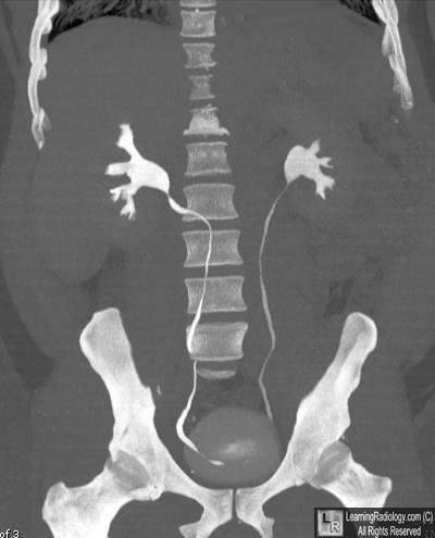
Coronal Reformatted CT Urogram
- Paget disease
- Bladder rupture
- Retrocaval ureter
- Retroperitoneal fibrosis
- Avascular necrosis
Additional Images-Axial CT image of abdomen
![]()
Additional Images

Axial CT image of abdomen
![]()
Answer:
3. Retrocaval ureter
More (Click Discussion Tab)
RetroCaval Ureter
General Considerations
- Also known as “circumcaval ureter”
- Abnormality in embryogenesis of IVC
- Results from abnormal persistence of right subcardinal vein positioned ventral to ureter in the definitive IVC
- Developing right ureter courses behind and medial to the IVC
- Incidence
- 0.07%
- Male to female ratio of 3:1
MORE . . .
.
This Week
37 year old male with hematuria |
Presented as a series of cards, this podcast asks some of the most common causes of neuroimaging findings and diseases making it ideal for a quick review. Can be used as either an audio only or audio/video podcast.; Complements Video Flashcard Podcasts 15, 21,25, 38, 42, 46 and 47. |
Some of the fundamentals of interpreting chest images |
The top diagnostic imaging diagnoses that all medical students should recognize according to the Alliance of Medical Student Educators in Radiology |
Recognizing normal and key abnormal intestinal gas patterns, free air and abdominal calcifications |
Recognizing the parameters that define a good chest x-ray; avoiding common pitfalls |
How to recognize the most common arthritides |
Now Open!
LearningRadiology
Named Magazine's
"25 Most Influential"
![]()
See Article on LearningRadiology
in August, 2010
RSNA News
| LearningRadiology.com |
is an award-winning educational website aimed primarily at medical students and radiology residents-in-training, containing lectures, handouts, images, Cases of the Week, archives of cases, quizzes, flashcards of differential diagnoses and “most commons” lists, primarily in the areas of chest, GI, GU cardiac, bone and neuroradiology. |
![]()